| [Previous] [Next] |
MRI provides excellent soft tissue contrast and can readily demonstrate and differentiate adjacent soft tissue structures, far superior to that possible with CT.
Everything is so beautifully seen on MRI that it is easier to discuss what isn't well seen. Air does not give off an MRI signal and produces a void on MRI, thus air containing structures such as the lung appear black. Likewise, MRI of the bowel is limited due to the presence of intestinal gas. Bowel peristalsis also causes motion artifact, limiting the utility of MRI in the evaluation of the bowel.
ABDOMEN
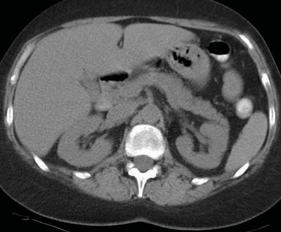
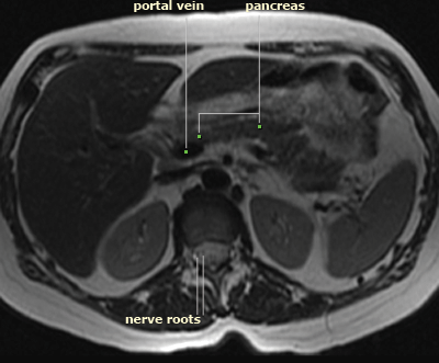
The images to the right were both obtained without iv contrast enhancement. The image on the top is a CT and the image on the bottom is a T2 weighted MRI. Note that on the CT all of the blood vessels and solid organs (liver, pancreas, spleen and kidneys) appear the same shade of gray. Note that on the MRI all of these structures are subtly different shades of gray, allowing us to distinguish them. Also note that the portal vein can be discretely seen from the adjacent pancreas on MRI, and individual nerve roots of the spinal canal are visible.
|